Lung Intelligence for COVID-19
COVID-19 Challenges
![]() Predicting progression & mortality risk to inform treatment decisions -- when to be aggressive with therapy?
Predicting progression & mortality risk to inform treatment decisions -- when to be aggressive with therapy?
![]() Understanding comorbidity to get a complete patient picture -- patients with COPD have a 5.9 fold higher risk of COVID progression.1
Understanding comorbidity to get a complete patient picture -- patients with COPD have a 5.9 fold higher risk of COVID progression.1
![]() Understanding complications and long lasting effects in recovering patients -- who is at risk of readmission & why is recovery stalling?
Understanding complications and long lasting effects in recovering patients -- who is at risk of readmission & why is recovery stalling?
VIDA's Lung Intelligence empowers physicians to address the above challenges
Quantitative Precision
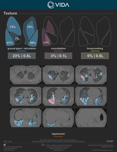
VIDA Insights provides quantitative measures and distribution of ground glass/reticulation, consolidation and honeycombing.
An overlay of these findings is also provided together with hyperlucent tissue, indicative of emphysema.
![]() Published works are indicating a primary imaging feature of COVID-19 to include ground-glass opacities with or without consolidation.2,3
Published works are indicating a primary imaging feature of COVID-19 to include ground-glass opacities with or without consolidation.2,3
![]() Quantitative lung consolidation is associated with the occurrence of major adverse hospital events for COVID-19 patients.4
Quantitative lung consolidation is associated with the occurrence of major adverse hospital events for COVID-19 patients.4

Texture Analysis, COVID-19 Case
Texture Visualization
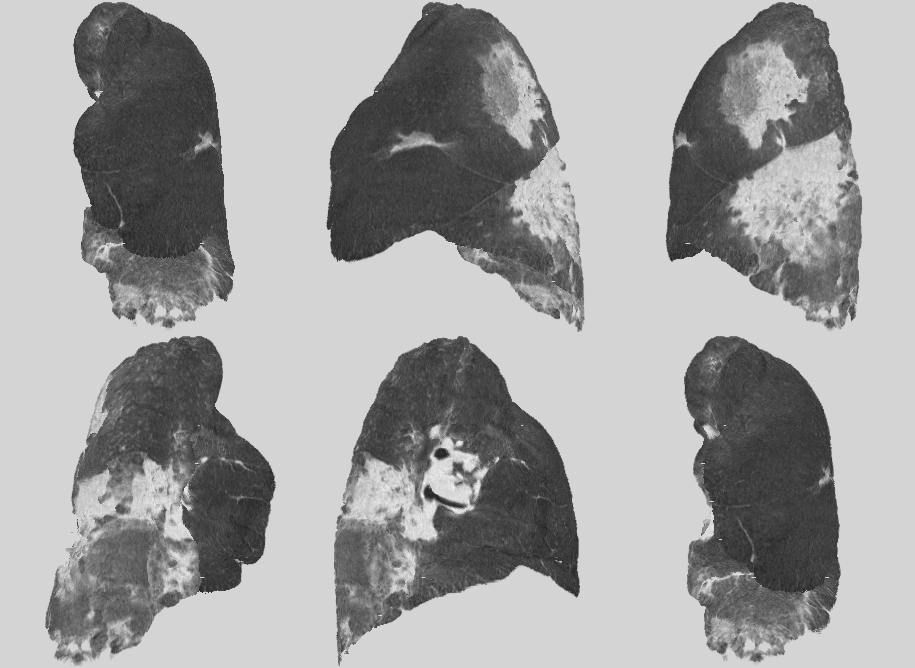
Subpleura View provides an impactful visualization of the 1mm space below the pleura, to quickly identify the presence or absence of textures in this space. This is especially useful in ILD and COVID-19 cases where these diseases often manifest.
Subpleura View is our latest visualization innovation, joining tMPR (topographic MPR) and others to give providers a modern way to see key imaging features. Subpleura View is patent pending.

Subpleura View, COVID-19 Case
Comorbid Assessment
![]() VIDA Insights also provides density analysis and tMPR visualizations.
VIDA Insights also provides density analysis and tMPR visualizations.
- Comorbid Assessment of underlying conditions, such as COPD, are key to understanding the increased vulnerability of disease progression with COVID-19 and/or complications associated with recovery. VIDA Insights provides:
Low attenuation (LAA%), a validated surrogate measure of emphysema. COVID-19 patients with COPD have a significantly higher risk of progression and readmission.5-7
Well-aerated lung (normal density) - lung parenchyma was found to be a strong predictor of intensive care unit (ICU) admission or death.8
-1.png?width=500&height=647&name=MicrosoftTeams-image%20(1)-1.png)
Density Analysis, COVID-19 Case


tMPR Visualizations, COVID-19 Case
VIDA Insights is clinically cleared in the US, EU, Australia & Canada. Texture and Subpleura View are available in the US and Canada under the public health emergency and available for research use in the EU and Australia.
References
1 -Wang, Bolin, Ruobao Li, Zhong Lu, and Yan Huang. “Does Comorbidity Increase the Risk of Patients with COVID-19: Evidence from Meta-Analysis.” Aging 12 (April 8, 2020). https://doi.org/10.18632/aging.103000.
2 - Colombi et al “Well-Aerated Lung on Admitting Chest CT to Predict Adverse Outcome in COVID-19 Pneumonia.” Radiology, April 17, 2020, 201433. https://doi.org/10.1148/radiol.2020201433.
3. Wang, Bolin, et al. “Does Comorbidity Increase the Risk of Patients with COVID-19: Evidence from Meta- Analysis.” Aging, vol. 12, Apr. 2020. PubMed, doi:10.18632/aging.103000
4- Sapienza, Lucas G., Karim Nasra, Vinícius F. Calsavara, Tania B. Little, Vrinda Narayana, and Eyad Abu-Isa. “Risk of In-Hospital Death Associated with Covid-19 Lung Consolidations on Chest Computed Tomography – A Novel Translational Approach Using a Radiation Oncology Contour Software.” European Journal of Radiology Open 8 (January 1, 2021): 100322. https://doi.org/10.1016/j.ejro.2021.100322.
5 - Schroeder J, et al. Relationships between airflow obstruction and quantitative CT measurements of emphysema, air trapping and airways in subjects with and without chronic obstructive pulmonary disease. AM J Roentgenol. 2013 Sept; 201(3).
6. Zach J, et al. Quantitative CT of the lungs and airways in healthy non-smoking adults. Invest. Radiology. 2012 Oct; 47(10).
7. Lowe K, et al. COPDGene 2019: Redefining the diagnosis of Chronic Obstructive Pulmonary Disease. JOPDF. 2019 Nov; 6(5)
8. Colombi et al “Well-Aerated Lung on Admitting Chest CT to Predict Adverse Outcome in COVID-19 Pneumonia.” Radiology, April 17, 2020, 201433. https://doi.org/10.1148/radiol.2020201433


